Hip Pain: Causes, Diagnosis, and Treatment

The hip joint bears the greatest load in the body. They are caused by the weight of walking, jumping, running, lifting and carrying heavy objects. Patients often experience hip pain. Orthopedic surgeons at specialized hospitals use modern diagnostic equipment to determine its cause. Doctors determine the extent of joint damage, which allows them to make an accurate diagnosis and develop the best treatment strategy.Professional doctors provide complex treatments for conditions that cause hip pain. The patient individually chooses an effective drug that affects the cause and mechanism of pain. Rehabilitation clinic experts provide rehabilitation treatment using the latest physical therapy procedures, physiotherapy and acupuncture. The presence of special simulators allows you to reduce the load on the joints during training.In treating hip pain, doctors from many medical fields are involved: endocrinologists, rheumatologists, orthopedists, physical therapists, chiropractors, acupuncturists. A multidisciplinary approach to treating hip pain can provide quick relief. Patients with hip pathology often require outside care.hip pain

reason

Hip pain is caused by the following pathological processes:
  • Tendonitis (inflammation of tendons);
  • muscle rupture;
  • Iliotibial band syndrome;
  • other local changes in surrounding tissues;
  • Systemic diseases (rheumatoid arthritis, polymyalgia).
Because the gluteus medius and minimus play a major role in hip abduction, injury to them can lead to hip pain. The gluteus medius and gluteus minimus tendons attach to the greater trochanter. If an inflammatory process occurs due to microtrauma caused by overloading, the patient will suffer from hip pain. Such diseases may be caused by infectious processes (tuberculosis), exercise or stereotypical occupational stress, or crystal deposition.

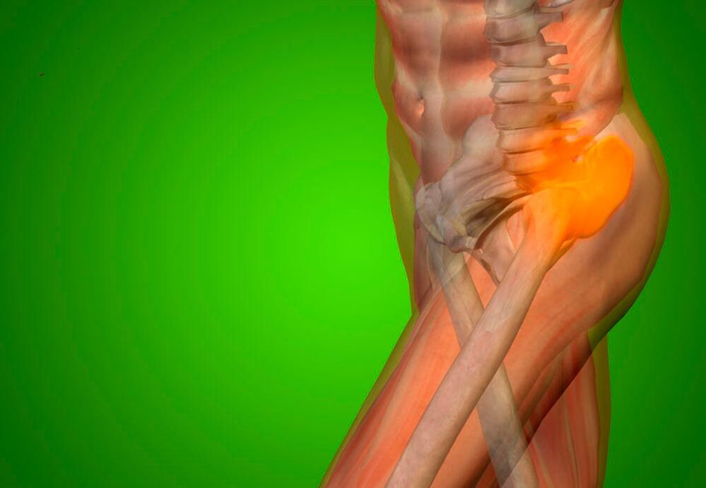
Hip pain is a symptom of:
  • Osteoarthrosis;
  • Nerve root syndrome;
  • Rheumatoid Arthritis;
  • cosita.
Hip pain can affect people who are overweight, have legs of different lengths, or have flat feet. Pain syndromes may occur after lower limb amputation or hip replacement. The patient complained of acute hip pain due to avascular necrosis of the femoral head and femoral neck fracture. Pain syndromes often occur with hip dysplasia (anatomical disorder). Acute pain in the hip joint that radiates down the leg can occur due to pinched nerves due to spinal disease, malignant bone tumors, and age-related changes.

Examination method

During the first consultation, the rheumatologist will conduct a thorough examination of the patient:
  1. Collect complaints and clarify the nature of hip pain;
  2. Obtain information about the course of the disease, onset of pain, progression of pain, and family and occupational factors that the patient believes contribute to the pain;
  3. External examination allows doctors to identify significant deviations from normal. To understand the nature of the pain and where it spreads, doctors ask patients to perform various movements of the hip joints of the lower limbs. Poor posture may indicate pathology in the hip joint;
  4. Palpation (feeling). Doctors can spot rheumatoid and rheumatic nodules, detect the exact location of pain during leg movement, and determine the moisture and temperature of the skin in the hip area.
Next, the doctor performs goniometry - an examination using a goniometer device. It allows you to determine your joint range of motion. The rheumatologist will then perform clinical and biological blood tests as well as a general urine test. The hospital's laboratory technicians use high-quality reagents and modern equipment to conduct research, which allows you to get accurate test results.

With inflammation of the hip joint, the number of white blood cells in the blood increases and the erythrocyte sedimentation rate increases. Increased levels of C-reactive protein in the serum indicate the inflammatory nature of the disease.

An immune blood test shows the presence of antinuclear antibodies in the blood of people with rheumatic inflammatory diseases. In patients with arthritis, the concentration of uric acid in the serum increases dramatically. Changes in the content of lysosomal enzymes (acid protease, acid phosphatase, cathepsin, deoxyribonuclease) in serum and synovial fluid of patients with rheumatism, psoriatic polyarthritis, rheumatism, and ankylosing spondylitis. In severe hip pathology, significant deviations from normal values are observed in urinalysis.

Doctor in clinic performs X-ray on patient with hip pain. Displayed when:
  • Chronic or acute pain in the hip joint at rest and during exercise;
  • Difficulty in moving lower limbs;
  • Swelling and discoloration of the skin in the hip area.
Doctors at the clinic use computed tomography scans to evaluate the bones involved in the formation of the hip joint. On computed tomography, radiologists detect changes in bone tissue structure, cartilage growth, and osteophytes.

Doctors use magnetic resonance imaging to evaluate the condition of the soft tissue surrounding the hip joint.

Radionucleotide research methods make it possible to identify pathologies using radiopharmacological drugs.

An ultrasound of the hip joint is performed to check for injuries, inflammatory diseases, rheumatism, and rheumatoid arthritis. The attending physician selects the research methods needed to determine the cause of hip pain individually in each case.

Differential diagnosis

Hip pain when walking is the main complaint that patients seek medical attention. It can be located in the joint area, or it can extend into the thigh, hip, or knee joint. If the hip joint hurts during exercise, the patient is forced to use crutches. Often, due to pain, mobility is limited when moving the hip joint, especially when rotating the leg outward and inward.

Pain in the hip, buttocks, and groin area is a symptom of aseptic necrosis of the femoral head. The disease is often associated with long-term use of hormonal drugs and alcohol abuse. As femoral head deformity progresses, hip joint movement is restricted. In the early stages of the pathological process, range of motion may be normal.

People with iliopectineal bursitis suffer from pain in the front of the hip joint and a clicking sound when moving the joint. It radiates to the thigh and is accompanied by paresthesias (tingling, burning, crawling sensations) due to compression of the femoral nerve. The patient experiences pain in the hip joint when bending or extending the lower limb. Pain can also be detected with deep palpation of the femoral triangle (the structure bounded by the inguinal ligament, the outer edge of the adductor longus muscle, and the inner edge of the sartorius muscle).

Pain on the outside of the hip joint is a sign of iliotibial band syndrome. There is a clicking sound during movement and pain on the outside of the knee joint, which worsens with movement.

Ross myalgia presents with burning pain in the anterolateral hip and thigh that worsens with walking and straightening the leg. Hip pain is associated with dysplasia. Over time, the patient develops a characteristic "duck" gait (he waddles from side to side as he walks).

hip pain

Hip pain is caused by coxarthrosis, a disease characterized by the degenerative process of the bones that form the joints. This disease more commonly affects older adults. As we age, the cartilage tissue in our joints loses elasticity, becomes thinner, and begins to wear away. When the load on the joint increases, the thin cartilage tissue is destroyed. The joint surfaces of bones rub against each other, causing aseptic inflammation.

Growths appear on the bones. They significantly limit joint movement. The joint surface becomes deformed, causing severe pain. Treatment of the disease depends on the severity of the joint damage. Doctors provide medication. If ineffective, endoprosthesis or palliative care is performed.

After determining the cause of hip pain, doctors begin treating the condition causing the pain syndrome. An expert committee meeting with the participation of professors, doctors and medical candidates, doctors of the highest rank, discussed cases of serious diseases in which patients suffered from hip pain.

treat

An important condition for successful treatment of conditions causing hip pain is the elimination of factors causing structural changes in the bone, cartilage and soft tissue of the joint area. For acute pain, hospital rheumatologists will prescribe nonsteroidal anti-inflammatory drugs. Through the use of topical treatments (topical gels and ointments, patches containing NSAIDs), patients' health improved significantly. They reduce pain in the hip joint during soft tissue inflammatory processes (tendonitis, bursitis, epicondylitis) after injury.

If this treatment is not effective enough, doctors may inject corticosteroids into the hip joint cavity. The joint space of the deformable hip joint is narrow and entry is difficult. Therefore, rheumatologists perform the surgery under X-ray control in specialized clinics. When inflammation of muscles and tendons causes pain, corticosteroids are injected into the tissues around the joints.

To improve cartilage condition and reduce hip pain, use a chondroprotectant. The course of treatment lasts several months. When the muscles involved in hip movement spasm, muscle relaxants are needed to reduce the tone of the skeletal muscles.

Medication is supplemented by physical therapy procedures. They are minor to hip pain. Due to the deeper location, physical therapy methods are less effective. The severity of hip joint pain is reduced after exposure to mid- and long-wave UV rays.

In the presence of inflammatory processes, high-intensity centimeter wave therapy, infrared laser therapy and low-intensity UHF therapy are performed. High-intensity high-frequency magnetic therapy, ozone therapy, and shock wave therapy stimulate tissue repair. The intensity of pain due to circulatory disorders and hip joint nutrition is reduced under the influence of various types of electrotherapy (exposure to electric current) and ultrasound.

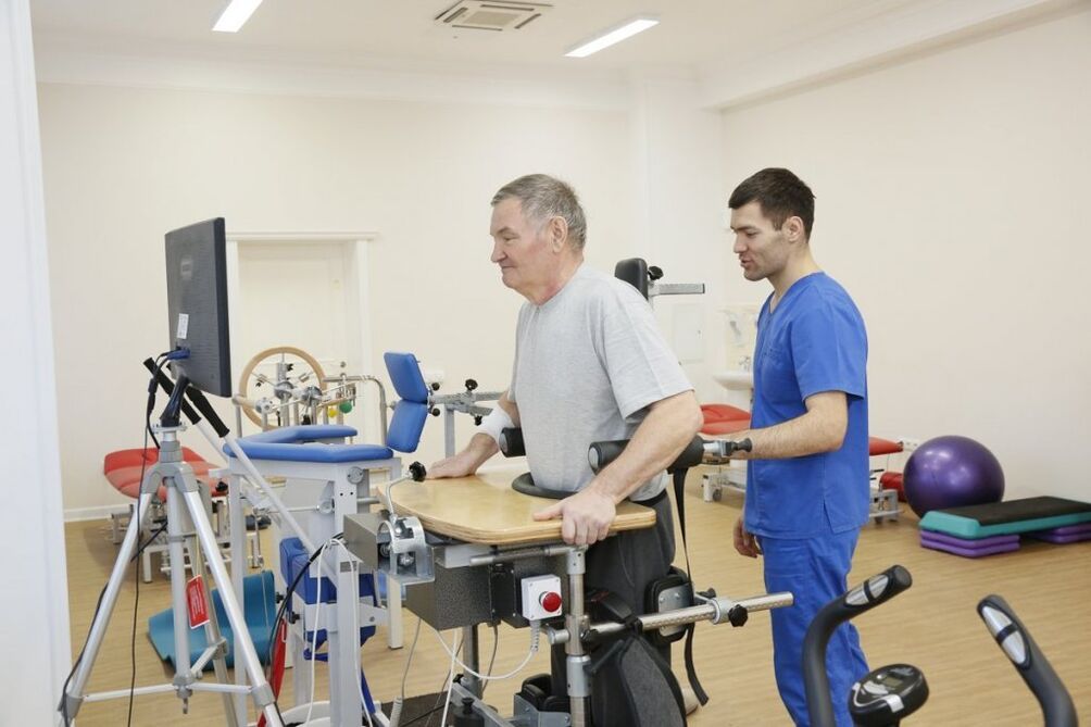
To reduce the load on the hip joint, rheumatologists recommend that patients use crutches when experiencing severe pain. After reducing the severity of the pain syndrome, the rehabilitation practitioner performs therapeutic exercises. An individual set of exercises is developed for each patient to quickly restore lower extremity function. When the structures involved in the formation of the hip joint are destroyed, the pain can be so severe that the only way to eliminate the pain is to replace the joint with an endoprosthesis.

Nonsteroidal anti-inflammatory drugs are used to relieve pain. Treatment depends on the condition affecting the hip joint. Patients are prescribed chondroprotectants to treat cartilage tissue damage. An orthopedic surgeon will prescribe effective treatments, diet, and exercises to improve circulation to the joints, restore cartilage tissue, and maintain joint mobility. In severe cases, joint replacement with an endoprosthesis is required, which can significantly improve quality of life and eliminate pain.
Treating Pelvic Pain with Exercise Therapy

exercise therapy treatment

Treating your hip joint using rehabilitative techniques allows you to maintain its mobility, improve blood circulation within the joint, and speed up the recovery of cartilage tissue. Rehabilitation specialists will select a set of physical therapy exercises based on the patient's joint disease. Rehabilitation sessions take place daily under the supervision of a coach. For rehabilitation treatment, special simulators are used and physical therapy programs are developed in conjunction with physical education.

What diseases cause joint pain

Right or left hip pain may be a sign of avascular necrosis of the femoral head. This disease occurs primarily in men and affects only one joint. Treatment involves eliminating pain, restoring blood supply to the joint area, normalizing the limb muscles, and maintaining joint function. Doctors prescribe painkillers, anti-inflammatory drugs, vitamins, physical therapy procedures and therapeutic exercises. Patients are advised to wear orthopedic shoes and use extra support when moving.The cause of hip pain may be a suppurative process. Primary septic arthritis occurs when there is a wound or injury and infectious agents enter the joint space. Secondary suppurative processes occur when septic or infectious agents enter the joint from surrounding tissues affected by the inflammatory process. For the treatment of septic arthritis, antibacterial treatment is performed by specialized specialists. If pus accumulates in the joint cavity, the hip joint is punctured, the pus is drained, and antibacterial agents are injected into the joint cavity.Bursitis is inflammation of the joint membrane. To relieve pain, doctors inject anti-inflammatory drugs and corticosteroids. If purulent inflammation is present, clean the cavity of the periarticular bursa. In severe cases, the joint capsule that has undergone irreversible changes is removed using surgical endoscopic techniques.In osteoporosis, femoral neck fractures frequently occur. Patients experience severe pain when moving the hip joint that radiates to the groin and inner thigh. Legs turn outward. Bruising and swelling in the hip area. In this case, treatment is performed by a professional orthopedic surgeon.Traumatic hip dislocation is associated with hip pain. The hip is reduced under general anesthesia. Congenital hip dislocation is diagnosed immediately after birth. It manifests as severe pain when the legs are spread and the knees are bent. Treatment is performed using special orthopedic structures.If you or a loved one has hip pain, you should not self-medicate. Seek professional medical attention immediately. Patients with acute pain usually need to stay in the clinic for at least a week. If the pain is not severe, the patient can have hip joint disease checked by a professional doctor and be treated at home in strict compliance with the regulations.